How to diagnose
If a horse is displaying signs indicative of navicular syndrome, the vet will first conduct a thorough lameness evaluation.
Following the initial evaluation there are additional methods of establishing whether navicular syndrome is a likely cause. These methods include nerve blocks as well as radiographs.
Examination & observation
The vet will first conduct a thorough visual lameness evaluation to establish if medical intervention is required.
The initial tests will typically begin with a thorough examination of both front feet and an observation of the horse walking and trotting in straight lines on both hard and soft surfaces to evaluate the severity of the condition.
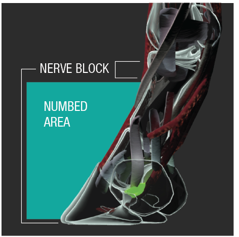
Nerve blocks & radiographs
Establishing the existence of pain
The vet will then apply hoof testers over the middle third of the frog of the hoof to see if pain is elicited when pressure is applied.
Confirming the source of the pain
By performing a palmar digital nerve block we can determine if the pain is coming from the navicular area; the horse will usually show significant improvement while trotting in hand and on the lunge line after desensitization.
Determining a prognosis
Grades of severity exist and a radiographic examination will better determine the condition of the navicular bone. Typical signs of the disease may vary from an asymetrical shape and poor definition between the cortex and medulla to the presence of large cyst-like lesions and new bone formation in advanced stages of the disease. Areas of increased resorption and formation are often seen in diseased navicular bones.